
Modules – Skills & Tools
Art of Microscopy & Life in a Drop of Water
Background Information
WHY YOU SHOULD CARE ABOUT… The Art of Microscopy
Using a microscope provides you with the unique opportunity to explore a world that’s hidden from our eyes. Most people in this world have no idea about the magic that lies in observing microscopic life. Microscopy skills help to determine a microorganism’s structure and characteristics that further aids in the investigation of unknown organisms that can possibly affect our daily lives. Microscopy skills are also essential as part of many careers including pathology laboratories, wastewater microbiology and many forms of research rely on a wide variety of microscopic techniques.
WHY YOU SHOULD CARE ABOUT… The Life in a Drop of Water
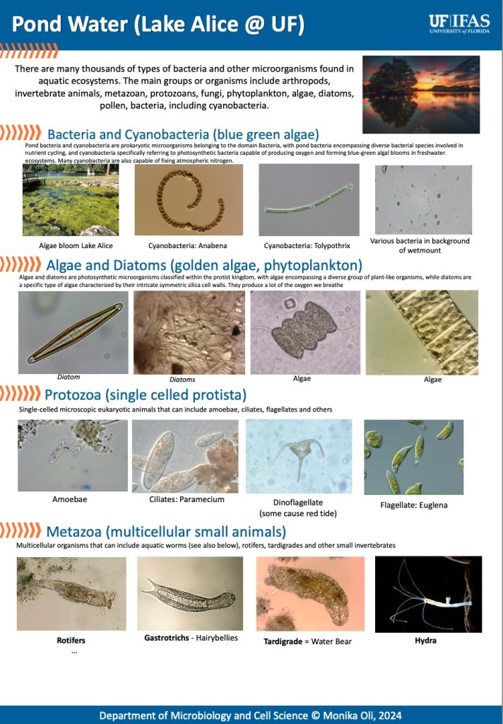
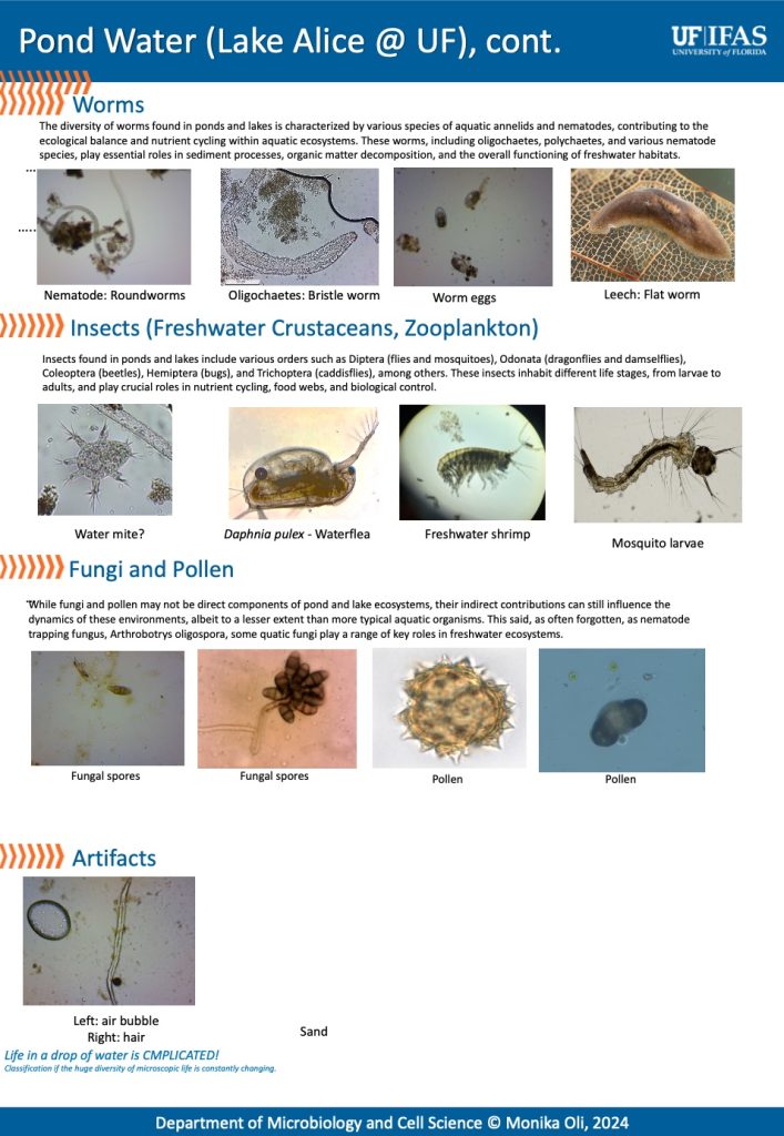
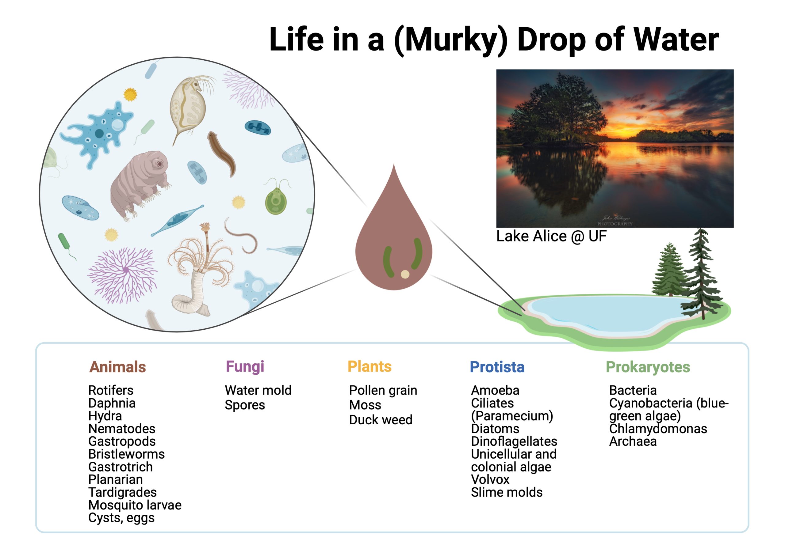
This simple observation experiment provides you with an opportunity to experience a whole new world of amazing microscopic organisms in a drop of water. You will find examples of many different organisms, including prokaryotes and eukaryotes, unicellular and multicellular organisms, protozoans, and algae. You can study the ecology of the ecosystem and examine and observe changes in the population composition of microbes such as amoebas, protozoa, rotifers (image), insect larvae, algae, and bacteria.
Learning Objectives
After completing this module, students will be able to:
1. Recall new vocabulary and definitions that pertain to this module.
2. Properly prepare and view specimens for examination using microscopy.
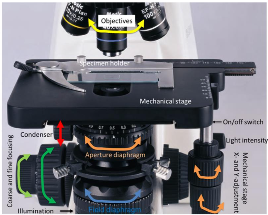
3. Identify the parts of a compound light microscope and know how to use it.
4. Measure the size of microscopic organisms and calculate the magnification of a specimen.
5. Recognize images taken with different microscopic techniques (light, dark field, differential interference contrast, fluorescence, TEM, SEM).
6. Explain the diversity and importance of protozoa and algae in our environment.
7. Recall the tardigrade – water bear and its mechanisms to survive under extreme conditions.
8. Connect microscopic observations and relate them to organismal evolution.
9. Describe the tree domains.
10. Sketch the difference between a prokaryote and a eukaryote.
History Connections
Microscope Terminology & Information
Microscope Vocabulary to know can be found here.
Classification of Organisms
The way we see and classify organisms has changed a lot since Linneaus original system, as depicted by the table below:
The most commonly referenced phylogenetic tree was established by Woese and is depicted below.
Virtual Lab Simulations
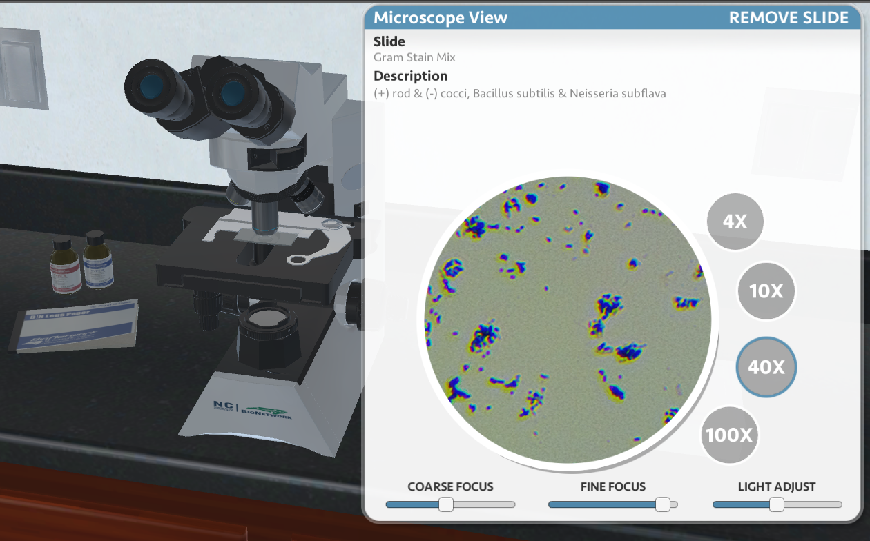
A Virtual Microscope Simulation:
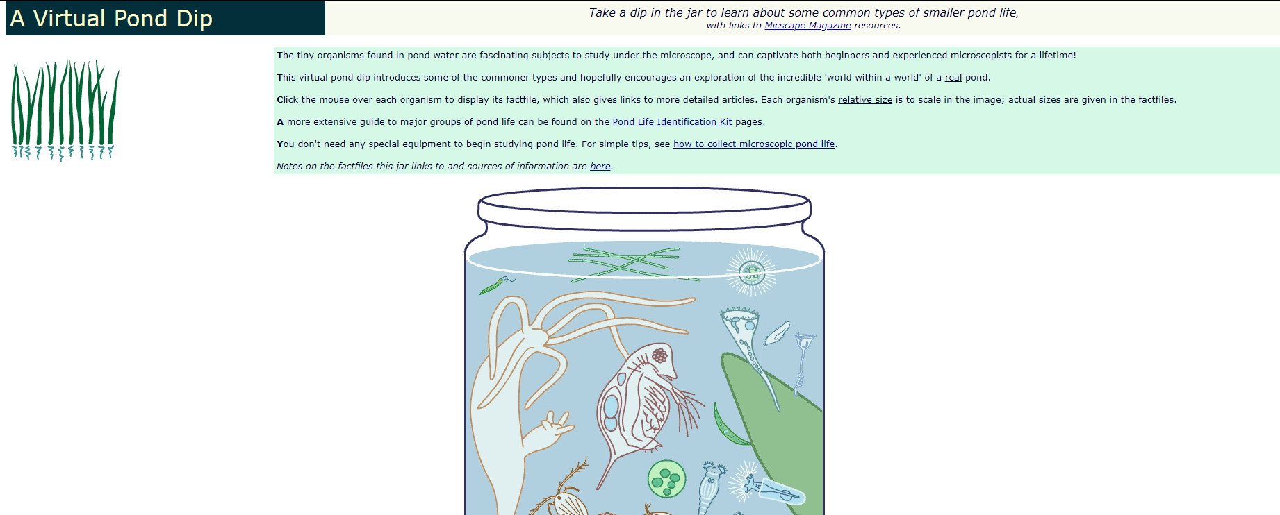
A Virtual Pond Dip Simulation:
Online Lab
Activity: Examining Life in a Drop of Water
-
Preparing a Wet Mount
-
Examine Your Wet Mount
-
Examine Prepared Slides
Lab Kit Materials for this Activity:
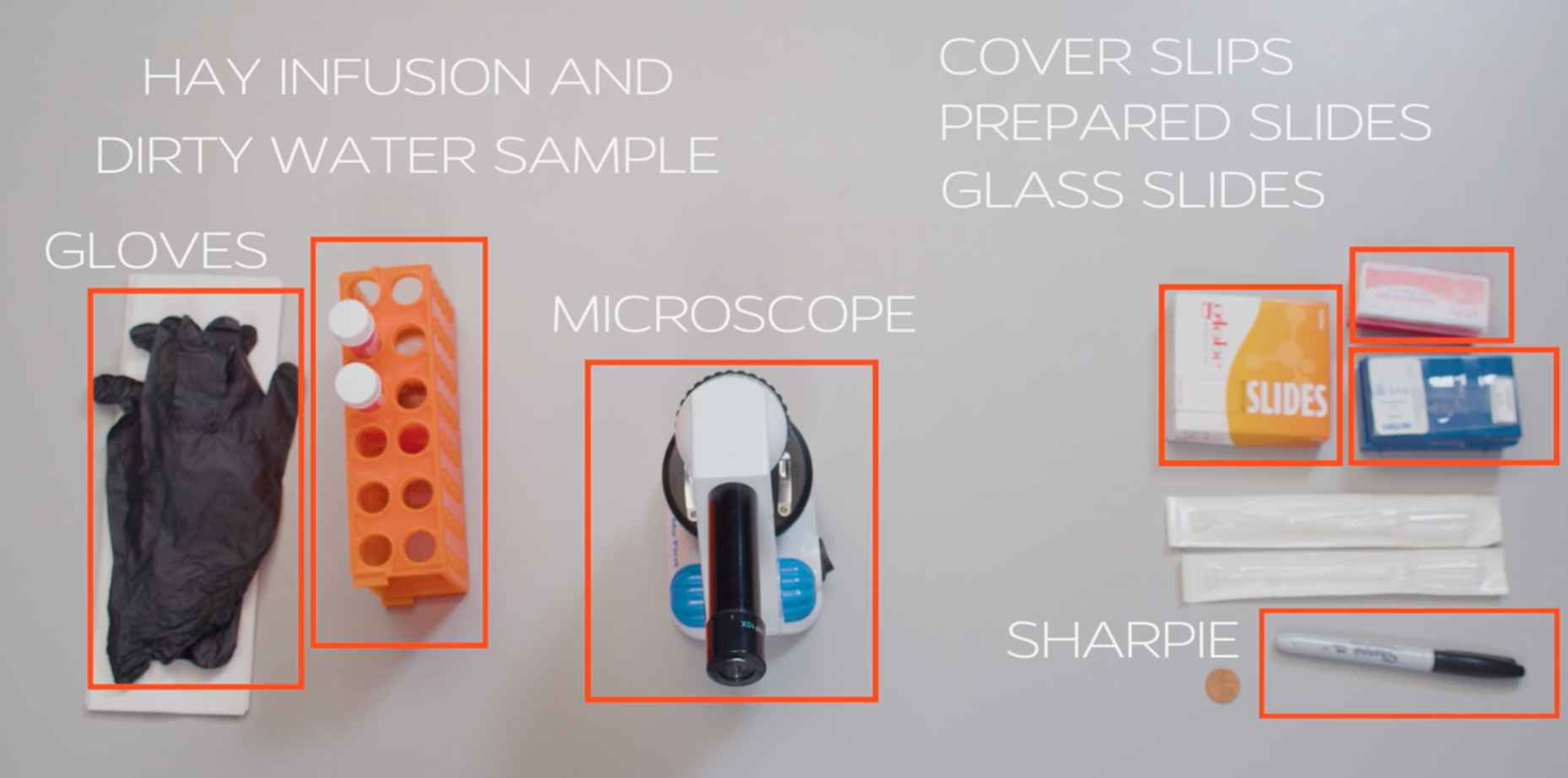
- Microscope
- Glass slides (1 box)
- Cover Slips (1 box)
- Prepared Slides (*Slides vary per student)
- Sharpie
- Water samples: Hay Infusion (from Week 1) and dirty water samples (student-provided)
-
- Other cool samples to examine:
Onion cells
Cheek swab
Anything else you are curious about!
- Other cool samples to examine:
-
Video Tutorials
Using your Microscope
Obtaining a Dirty Water Sample
Preparing & Examining a Wet Mount
PhyloT Tutorial
Procedure
Preparing a Wet Mount
- Using a transfer pipette add a small drop (20 µl) of water sample (from the hay infusion created in the Microbial Ecology module) to a microscope slide. Make sure you don’t just take water but look at samples from the top, middle and bottom of the sample. Most microorganisms will be where there is food, i.e., plant material
- Place the edge of the microscope coverslip on the microscope slide so that it slightly touches the edge of the sample.
- Lower the microscope coverslip over the sample.
- Place the microscope slide with the sample on the stage of the microscope and secure in place with the stage clips.
- Move the slide to the center directly over the light source. Use the bottom light source.
- Use the 4x and 10x objective first
- Observe various organisms.
- Repeat these procedures for all 3 water samples.
Examine your Wet Mount
- Using the 4x objective lens, focus on the hay infusion sample.
- Change to the 10x objective lens. Adjust both the focus and slide if necessary.
- Change to the 40x objective lens. Adjust both the focus and slide if necessary.
- Put your phone’s camera lens up to the eyepiece of the microscope to get pictures of the magnified image
- Identify protists and their movement, shapes and notable characteristics
- Scroll through the resources below and any online resources to identify microorganisms that you find in your water samples.
- Examine your box of prepared slides. Feel free to further explore this module by looking up images of the following slides if they are not included in your kit:
- Frog blood
- Trypanosoma cruzi or brucei in blood
- Malaria
- Sickle cell anemia
Results
Document the following for your ELN:
Hay-Infusion:
Examine a drop from the hay infusion once a week for 4-5 weeks and fill out the table below in your ELN. Indicate whether you see the different organisms each week and how the types of microbe change. Try to identify as many of the organisms as you can and track their population changes over time.
| Organism | Week 1 | Week 2 | Week 3 | Week 4 | Week 5 | Week 6 |
Life in a Drop of Water:
Record in your ELN any organisms seen in your dirty water samples along with their identification, photo, and microscope magnification. Also, add any organisms you identify onto Phyto-T to make a phylogenetic tree.
Prepared Slides:
Discuss which slides were included in your kit and what you see (NOTE: Provided slides will be different for each student). Describe which objective setting you use to view each sample.
In-Person Lab
Art of Microscopy In-Person Procedure:
Life in a Drop of Water In-Person Procedure:
Video Tutorials
How To Use a Microscope
How To Kohler a Microscope
Preparing a Wet Mount
Resources
Art of Microscopy
- Kohlering (Aligning) a microscope tutorial.
- Info about different microscopes and tutorials by Nikon Microscopy U
- Microscope Information: about microscopes, images, and advice
Life in a Drop of Water
